
© elfruler 2025
References are found at the bottom of the page.
Several statements circulating in the eagle-cam watching world assert that saliva and nasal fluids passed from a breeding parent to a new hatchling provide important benefits, including digestive enzymes, antibodies, and moisture to enable the chick to swallow food.
I know of only a handful of comments in publications about raptors that refer to saliva passing from parent to chick, none of them supported by citations of scientific evidence. For instance, in his book Birds of Prey (1995, targeted mainly to falconers), Fox remarks that “females feeding young chicks drool large quantities of liquid onto the food for the chick to ingest and it is not clear if this fluid is salivary or nasal, or both, in origin” – or, I would add, from the food itself (emphasis added). Wiemeyer 1981 comments in a report on breeding by captive Bald Eagles that “when the eaglets were less than 1 week old a saliva-like fluid was seen dripping from the tip of the upper mandible of the adult feeding the young, thereby providing extra moisture and possibly digestive enzymes to the young with the pieces of food” (emphasis added).
To learn whether any of the claims are valid, I took a deep dive into peer-reviewed research on the biology of avian saliva, nasal secretions, and digestive and immune systems, as well as embryological development of a chick.
What is the make-up of saliva and nasal fluids?
As in all vertebrates, saliva in birds is made up primarily of mucus, a mixture of water and the protein mucin. Mucus keeps the oral cavity lubricated and moistens food to make it easier to swallow. Both mucus and the membrane whose cells produce it serve as barriers to toxins and pathogens that may enter the mouth. The nasal cavity just inside the nostrils (or nares) also is lined by a membrane that secretes mucus, which helps trap incoming debris and can protect against foreign agents. It is normal for excess mucus to drip out of the nares or be expelled in the form of a sneeze.
Do new hatchlings produce their own saliva and nasal fluids?
An eaglet embryo’s salivary glands begin to form toward the end of the second week of development, and they are developed and functioning by the time the eaglet hatches. The mucous membranes in the oral and nasal cavities are producing mucus by the middle of the third week of embryonic development. Embedded in the nasal membrane are nerve endings which convey smell signals to the bird’s olfactory bulb, and these neurons start working as the membrane begins functioning. This means that the embryos can learn to distinguish smells before hatch, even before the internal pip of the air sac. (Recent research shows that most birds have a stronger sense of smell than previously believed.)
Do hatchlings have difficulty swallowing?
Anyone watching the first feeding of a hatchling eaglet, which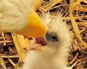 can happen within 2 or 3 hours of hatch, knows that it has no problem gulping down small, solid chunks of meat offered by a parent. A hatchling’s tongue already has baby-sized rear-pointed papillae extending from the back of the tongue, which push the food back toward the esophageal opening.
And clearly the reflexive movement that sends the food down the esophagus is functioning. In this slo-mo video (click on the image), note that the parent turns its beak sideways to make it easier for the chick to access the food. This position allows some liquid to drip out of the side of the beak and into the nest – not into the chick’s mouth.
Studies have shown that bird embryos can swallow even before hatching. About two-thirds of the way through the incubation period they begin swallowing some of the amniotic fluid in which they are bathed.
Are there digestive enzymes in saliva or nasal secretions?
In some species, yes, but as far as I can learn, none have been detected in Bald Eagles. The only digestive enzymes securely identified in avian saliva are those that target sugars and complex carbohydrates and break them down into digestible form. The most common such enzyme, amylase, has been found in the saliva of a few species that eat seeds, grasses, insects, and fruits, a diet that is plentiful in sugars and carbs. But the diet of carnivores like Bald Eagles is not, so this kind of enzyme is unnecessary, and scientists have found little to no amylase anywhere in their digestive tracts.
How well are new hatchlings equipped to digest food?
A further dive into studies of embryology casts light on a new hatchling’s capacity to digest food and to fight off infection, needing no extra help from a parent’s saliva. An eaglet’s digestive system is fully functional by the time it emerges from the egg, complete with digestive enzymes. This is evident in its expulsion of wastes about 12 hours after its first feeding, when its system has extracted all the usable nutrients from the food. (click on the image for video)
But long before an eaglet hatches, digestive enzymes are produced in the yolk sac membrane. These break down the lipids, proteins, and carbohydrates in the yolk to supply the embryo with the energy it needs to grow. The digested nutrients are absorbed into the blood vessels that are spread across the membrane, and the capillaries convey the food into the embryo’s intestinal cells. These enzymes increase throughout embryonic development, and the intestine itself grows rapidly as hatch nears.
And, as is well known, just prior to hatch a chick absorbs what is left of the yolk sac, so its nutrients feed the chick, and its digestive enzymes help it process new food during its first 24-48 hours out of the shell.
Does saliva contain antibodies?
I found no evidence in the scholarly literature of any appreciable amount of antibodies in the saliva of birds. The only such assertion pertaining to Bald Eagles that I have seen is in Nielsen 1991, a picture-book about a breeding season of a pair of Bald Eagles, which lists no references. The idea may have origins in the phenomenon of “crop milk” that pigeons and doves feed to their hatchlings. Crop milk, as the name implies, consists of fatty cells that are shed from the lining of the parent’s crop, mixed with a healthy proportion of water. Rich in lipids and proteins and some antibodies, it is regurgitated and fed to the chicks during the first few days after hatch. (And, of course, it is not strictly “milk” as it is not secreted by a mammary gland.) Eagles do not produce crop milk.
Do new hatchlings have any immunity against infection on their own?
To answer that question, it’s useful to survey how immunity works in birds. Like all vertebrates, birds are protected by two types of immunity: innate immunity and adaptive immunity.
Innate immunity is the natural, non-specific, protection that is provided by cells throughout the body. Agents of innate immunity do not target specific pathogens. They include physical barriers against penetration by harmful microbes, including skin and scales, feathers, and membranes in the respiratory and digestive tracts. Membranes are very effective in thwarting infection by helping prevent absorption of toxins and pathogens. Mucous membranes host macrophages, white blood cells that can ingest foreign cells and break them down, especially in the intestine. Mucus can trap infectious organisms and transport them from the body in a sneeze, phlegm, excretion, or sputum (which can be swallowed and expelled via the digestive system). Beneficial microorganisms in the skin, mucous membranes, and especially the digestive tract can neutralize bacteria & viruses. Defensive peptides can disable infectious agents.
By the time an eaglet hatches, its innate protections are well developed. Mucous membranes begin to develop during the second and third weeks of embryological development, and intestinal cells are producing mucin by the end of the embryonic period. Early forms of macrophages develop in the yolk sac and are functional in the digestive system by the time a chick hatches. Infection-fighting peptides appear as early as the second week of embryonic development. The developing embryo receives beneficial microorganisms from its mother during egg formation, and during brooding more such microbiota easily transfer to the hatchling from both parents.
Adaptive immunity is provided by antibodies, proteins that target specific pathogens and adapt to retain a memory of them for future defensive action. Antibodies are produced by white blood cells, or lymphocytes, named B cells for the organ in birds where they mature, the Bursa of Fabricius (after the 17th-century scientist who discovered it). This organ is now commonly referred to as the cloacal bursa, as it is a small sac growing out of the wall of the cloaca. B cells and antibodies “learn” the pathogen so they can recognize it if it invades again – what we call “immunity.”
Mature B cells from the bursa populate lymphatic tissues throughout the body, including mucous membranes, the spleen, areas of the intestine, the bronchi in the respiratory tract, skin, and the nasal cavity. When a B cell encounters a pathogen, it is activated to multiply and to produce increasing numbers of antibodies that can bind to that pathogen and destroy or disable it. The action of B cells is reinforced by lymphocytes called T cells, which mature in the Thymus gland and also populate lymphatic tissues throughout the body.
But long before hatch, a bird embryo has a good supply of antibodies provided by its mother. She deposits antibodies into the yolk while it is forming in her ovary, along with fats and proteins and other nutrients. And when the egg moves down the oviduct, more maternal antibodies are transferred into the albumen. Antibodies in the yolk enter the embryo’s blood stream beginning a little over halfway through incubation. At about the same time, the embryo swallows albumen and its antibodies.
The embryo’s own adaptive immune system takes a little longer than innate protections to mature. The cloacal bursa begins to grow in an embryo’s second week, and lymphocyte stem cells appear there in the last third of the incubation period, where they begin to proliferate and to develop antibody-producing capability. Lymphocytes enter the embryo’s blood stream in the third week of incubation and begin to migrate to other lymphatic tissues about a week before hatch, slowly at first, then increasing till hatch. By that time, most of the lymphocytes in the bursa are mature and ready to produce antibodies.
Stem cells of the thymus start to develop in the embryo by about the second week, and the organ is well developed by the fourth week. Lymphocyte stem cells from the spleen and liver travel to the thymus in waves between about the middle of the second week to the fifth week, where they mature into antibody-capable T cells. In the last week of incubation they begin to migrate out to peripheral lymphatic tissues where they can begin to detect foreign cells.
The transfer of maternal antibodies from the yolk into the embryo increases significantly during the last 3-5 days of incubation and are at a high level in the chick at hatch, but then they begin to decrease. The maternal antibodies are specific for pathogens encountered by the mother during yolk deposition and egg formation in her oviduct, but a chick will encounter new pathogens in its wider environment. So its own antibody-producing capability ratchets up at an increasing pace after hatch. The bursa increases in size quickly and produces an increasingly diverse repertoire of B cells and antibodies, especially in response to infectious agents in the environment, and their dispersion to other lymphatic tissues speeds up. The Harderian gland, a small organ behind the eye in the optical orbit, is functional by the time the chick hatches. It secretes fluid that lubricates the cornea of the eyes & the nictitating membrane, and the secretion contains lymphocytes and antibodies that help protect the hatchling’s precious eyes from infection.
The cloacal bursa remains active while the eaglet continues to grow, but it regresses by the time the bird is sexually mature, in response to increasing gonadal hormones. The thymus gland follows the body’s annual cycle, reaching a peak during molt and shrinking in fall and winter until the next breeding cycle has begun. Like the cloacal bursa, the thymus begins to atrophy later in the bird’s life. Lymphocytes that have moved to other lymphatic tissues continue to divide and produce antibodies in response to pathogens throughout a bird’s life.
To sum up
- There is no scientific evidence to support assertions that a Bald Eagle’s saliva or nasal fluids contain digestive enzymes or antibodies to transfer to hatchlings during feeding.
- By the time it hatches, an eaglet has been producing saliva and nasal fluids on its own.
- By the time it hatches, an eaglet has already been swallowing fluids inside the egg.
- During embryonic development the yolk sac membrane produces digestive enzymes to process the nutrients in the yolk and pass them into the blood stream of the developing embryo.
- By the time it hatches, a chick’s digestive system, including digestive enzymes, is fully functional.
- A breeding female deposits some of her antibodies into the yolk as it develops in her ovary, which are transferred into the embryo’s blood stream, and they continue to protect the eaglet from infection after it hatches.
- As the egg is forming in the oviduct, the female deposits some of her antibodies into the albumen, which is swallowed by the embryo, and these continue to protect the eaglet from infection after it hatches.
- By the time it hatches, a chick’s innate immune system (barriers, mucous membranes, macrophages, peptides, beneficial microorganisms) is in place and can protect against many potential infections.
- After hatching the chick’s adaptive immune system develops quickly and produces antibodies to protect against new pathogens encountered in the environment.
REFERENCES
Apanius, V. 1998. The immune system. In J.M. Starck and R.E. Ricklefs (eds.), Avian Growth and Development: Evolution within the Altricial-Precocial Spectrum. Oxford University Press: New York and Oxford: 203-222.
Ballou, A.L., R.A. Ali, M.A. Mendoza, J.C. Ellis, H.M. Hassan, W.J. Croom, and M.D. Koci. 2016. Development of the chick microbiome: how early exposure influences future microbial diversity. Frontiers in Veterinary Science 3.
Bhattacharya, S., and K.C. Ghose. 1971. Influence of food on the amylase system in birds. Comparative Biochemistry & Physiology 40B: 317-320.
Broom, L.J., and M.H. Kogut. 2018. The role of the gut microbiome in shaping the immune system of chickens. Veterinary Immunology and Immunopathology 204: 44-51.
Christ, B., and M. Scaal. 2007. Embryogenesis and development. In B.J.M. Jamieson (ed.), Reproductive Biology and Phylogeny of Birds, Part B: Sexual Selection, Behavior, Conservation, Embryology, Genetics. Enfield, NH: Science Publishers. 401-478.
Clark, L., and C.A. Smeraski. 2022. Chemesthesis and olfaction. In C.G. Scanes (ed.), Sturkie’s Avian Physiology. 7th ed. San Diego, CA: Academic Press. 179-203.
Davison, F. 2022. The importance of the avian immune system and its unique features. In B. Kaspers, K.A. Schat, T.A. Göbel and L. Vervelde (eds.), Avian immunology. London: Elsevier. 1-9.
Denbow, D.M. 2015. Gastrointestinal anatomy and physiology. In C.G. Scanes (ed.), Sturkie’s Avian Physiology. 6th ed. San Diego, CA: Academic Press. 337-366.
Ding, J., R. Dai, L. Yang, C. He, K. Xu, S. Liu, W. Zhao, L. Xiao, L. Luo, Y. Zhang, and H. Meng. 2917. Inheritance and establishment of gut microbiota in chickens. Frontiers in Microbiology 8.
El-Bakry, A.M., and S.-i. Iwasaki. 2014. Ultrastructure and histochemical study of the lingual salivary glands of some bird species. Pakistan Journal of Zoology 46: 553-559.
Evans, H.E. 2016. Bird physiology. In I.J. Lovette and J.W. Fitzpatrick (eds.), Handbook of Bird Biology. 3rd ed. Cornell, NY: Cornell University Press. 215-262.
Evans, H.E., and J.B. Heiser. 2004. What’s inside: anatomy and physiology. In S. Podulka, J. Ronald W. Rohrbaugh and R. Bonney (eds.), Handbook of Bird Biology. 2nd ed. Ithaca, NY: Cornell Lab of Ornithology. 4-1 – 4-162.
Yvernogeau, L., N. Nagy, D. Dunon, C. Robin, and T. Jaffredo. 2022. Development of the avian hematopoietic and immune systems. In B. Kaspers, K.A. Schat, T.A. Göbel and L. Vervelde (eds.), Avian immunology. London: Elsevier. 45-69.
Fentzloff, C. 1984. Breeding, artificial incubation and release of White-tailed sea eagles Haliaeetus albicilla. International Zoo Yearbook 23: 18-35.
Fisher, H. 1972. The nutrition of birds. In D.S. Farner, J.R. King and K.C. Parkes (eds.), Avian Biology. New York and London: Academic Press. 431-469.
Fox, N. 1995. Understanding the Bird of Prey. Hancock House Publishers: Blaine, WA; Surrey, BC.
Gabrielli, M.G., and D. Tomassoni. 2014. Carbonic anhydrase in minor salivary glands of quail: histochemistry versis immunohistochemistry. Journal of Enzyme Inhibition and Medicinal Chemistry 29: 87-91.
Ganchrow, D., and J.R. Ganchrow. 1985. Number and distribution of taste buds in the oral cavity of hatchling chicks. Physiology & Behavior 34: 889-894.
Glick, B. 2000. Immunophysiology. In G.C. Whittow (ed.), Sturkie’s avian physiology. 5th ed. San Diego, CA: Academic Press. 657-670.
Grindstaff, J.L., E.D. Brodie III, and E.D. Ketterson. 2003. Immune function across generations: integrating mechanism and evolutionary process in maternal antibody transmission. Proceedings of the Royal Society B: Biological Sciences 270: 2309-2319.
Hamilton, H.L. (ed.). 1952. Lillie’s Development of the Chick: An introduction to embryology. New York: Henry Holt & Company.
Kaiser, P., and A. Balic. 2015. The avian immune system. In C.G. Scanes (ed.), Sturkie’s Avian Physiology. 6th ed. San Diego, CA: Academic Press. 403-418.
Kaspers, B., K.A. Schat, T.A. Göbel, and L. Vervelde (eds.). 2022. Avian immunology. London: Elsevier.
Kaspers, B., I. Schranner, and U. Lösch. 1991. Distribution of immunoglobulins during embryogenesis in the chicken. Journal of Veterinary Medicine A 38: 73-79.
Kaspers, B., H. Bondi, and T.W.F. Göbel. 1996. Transfer of IgA from albumen into the yolk sac during embryonic development in the chicken. Journal of Veterinary Medicine A 43: 225-231.
Kincade, P.W., and M.D. Cooper. 1971. Development and distribution of immunoglobulin-containing cells in the chicken. The Journal of Immunology 106: 371-382.
Kogut, M.H. 2018. The effect of microbiome modulation on the intestinal health of poultry. Animal Feed Science and Technology 250: 32-40.
Kogut, M.H. 2022. Impact of the gut microbiota on the immune system. In B. Kaspers, K.A. Schat, T.A. Göbel and L. Vervelde (eds.), Avian immunology. London: Elsevier. 353-364.
Kogut, M.H. 2022. Immunophysiology of the avian immune system. In C.G. Scanes (ed.), Sturkie’s Avian Physiology. 7th ed. San Diego, CA: Academic Press. 591-610.
McWilliams, S., E. Adkins-Regan, and C. Vleck. 2016. Bird physiology. In I.J. Lovette and J.W. Fitzpatrick (eds.), Handbook of Bird Biology. 3rd ed. Cornell, NY: Cornell University. 215-262.
Murphy, J.B. 1914. Studies in tissue specificity. II. The ultimate fate of mammalian tissue implanted in the chick embryo. J. Exp. Med. 19, 181-186 (ref in Davison 2014)
Nagy, N., I. Oláh, and L. Vervelde. 2022. Structure of the avian lymphoid system. In B. Kaspers, K.A. Schat, T.A. Göbel and L. Vervelde (eds.), Avian immunology. London: Elsevier. 11-44.
Nicknafs, S., M. Navarro, E.R. Schneider, and E. Roura. 2023. The avian taste system. Frontiers in Physiology 14.
Nielsen, S. 1991. A season with eagles. Voyageur Press: Stillwater, MN, U.S.A.
Patten, B.M. 1971. Early embryology of the chick. 5th ed. McGraw-Hill Book Company: New York.
Powell, F.L. 2015. Respiration. In C.G. Scanes (ed.), Sturkie’s Avian Physiology. 6th ed. San Diego, CA: Academic Press. 301-336.
Proszkowiec-Weglarz, M. 2022. Gastrointestinal anatomy and physiology. In C.G. Scanes (ed.), Sturkie’s Avian Physiology. 7th ed. San Diego, CA: Academic Press. 485-527.
Réhault-Godbert, S., M. Huncke, R. Guibiraba, N. Guoyot, and J. Gautron. 2022. Innate defenses of the avian egg. In B. Kaspers, K.A. Schat, T.A. Göbel and L. Vervelde (eds.), Avian immunology. London: Elsevier. 365-386.
Ricklefs, R.E., and J.M. Starck. 1998. Embryonic Growth and Development. In J.M. Starck and R.E. Ricklefs (eds.), Avian Growth and Development: Evolution within the Altricial-Precocial Spectrum. Oxford University Press: New York and Oxford: 31-58.
Ricklefs, R.E., and J.M. Starck. 1998. The Evolution of the Developmental Mode in Birds. In J.M. Starck and R.E. Ricklefs (eds.), Avian Growth and Development: Evolution within the Altricial-Precocial Spectrum. Oxford University Press: New York and Oxford: 366-380.
Samar, M.E., R.E. Ávila, S.P. De Fabro, v. Porfirio, F.J. Esteban, J.A. Pedrosa, and M.A. Peinado. 1999. Histochemical study of Magellanic Penguin (Spheniscus magellanicus) minor salivary glands during postnatal growth. The Anatomical Record 254: 298-306.
Samar, M.E., R.E. Ávila, F.J. Esteban, L. Olmedo, L. Dettin, A. Massone, J.A. Pedrosa, and M.A. Peinado. 2002. Histochemical and ultrastructural study of the chicken salivary palatine glands. Acta Histochemica 104: 199-207.
Starck, J.M., and R.E. Ricklefs, eds. 1998. Avian growth and development: evolution within the altricial-precocial spectrum. Oxford University Press: New York.
Starck, J.M., and R.E. Ricklefs. 1998. Embryonic Growth Rate Data Set. In J.M. Starck and R.E. Ricklefs (eds.), Avian Growth and Development: Evolution within the Altricial-Precocial Spectrum. Oxford University Press: New York and Oxford: 381-415.
Vince, M.A. 1977. Taste sensitivity in the embryo of the domestic fowl. Animal Behaviour 25: 797-805.
Wiemeyer, S.N. 1981. Captive Propagation of Bald Eagles at Patuxent Wildlife Research Center and Introductions into the Wild, 1976-80. Journal of Raptor Research 15: 68-82.
Wong, E.A. 2022. Functional properties of avian intestinal cells. In C.G. Scanes (ed.), Sturkie’s Avian Physiology. 7th ed. San Diego, CA: Academic Press. 529-548.
Ziswiler, V., and D.S. Farner. 1972. Digestion and the digestive system. In D.S. Farner, J.R. King and K.C. Parkes (eds.), Avian Biology. New York and London: Academic Press. 343-430.
You must be logged in to post a comment.